The invention of the x-ray machine marked a significant milestone in the medical world. On this day in 1896, scientist Henry Louis Smith formally exhibited the first x-ray machine. However, when he unveiled it, people were confused and unable to comprehend what he was trying to show them. It was only months later that German physics professor Wilhelm Röntgen discovered x-rays while conducting experiments with Lenard and Crookes tubes, leading to further research in the field.

The simplicity of an x-ray machine, such as those used in doctor’s or dentist’s offices, may come as a surprise. Essentially, it produces a controlled beam of radiation that is used to create an image of the inside of the body. Inside the machine is an x-ray tube, which contains an electron gun that shoots high-energy electrons at a target made of heavy atoms like tungsten.
In today’s world, getting an x-ray done is a relatively quick and easy process. However, the first x-ray machine required individuals to sit in front of it for over an hour to capture the necessary images. Additionally, the radiation dose applied to the subject was approximately 1,500 times higher than what is typically used in modern x-rays.
The introduction of the x-ray machine revolutionized medical diagnostics. Doctors and medical professionals could now easily visualize and identify even the smallest abnormalities within the body, aiding in accurate diagnoses and treatment plans. The ability to capture detailed images without invasive procedures allowed for a non-invasive approach, significantly reducing patient discomfort and improving overall healthcare outcomes.
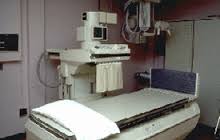
Since the invention of the x-ray machine, it has become an indispensable tool in the medical field. It has evolved over the years, with advancements in technology leading to the development of more efficient and safer machines. Today, x-rays are used to diagnose various conditions, including bone fractures, lung infections, and even dental issues.
The impact of the x-ray machine on medical science cannot be overstated. Its ability to provide detailed images of the internal structures of the body has transformed healthcare practices and saved countless lives. Henry Louis Smith and Wilhelm Röntgen’s contributions to the field of radiology have paved the way for numerous advancements in medical imaging, benefiting patients worldwide.
#Hindi
एक्स-रे मशीन के आविष्कार ने चिकित्सा जगत में एक महत्वपूर्ण मील का पत्थर स्थापित किया। 1896 में, वैज्ञानिक हेनरी लुईस स्मिथ ने पहली बार एक्स-रे मशीन का प्रदर्शन किया। जब उन्होंने इसे दिखाया, तो लोग भ्रमित हो गए और समझ नहीं पाए कि वह क्या दिखाना चाह रहे थे। कुछ महीने बाद, जर्मनी के भौतिकी प्रोफेसर विल्हेम रॉन्टजन ने लीनार्ड और क्रुक्स ट्यूब का प्रयोग करते हुए एक्स-रे की खोज की, जिससे इस क्षेत्र में और अधिक शोध हुए।
एक्स-रे मशीन की सरलता देखकर हैरानी हो सकती है, जैसे डॉक्टर या दंत चिकित्सक के कार्यालय में उपयोग होने वाली मशीनें। मूल रूप से, यह नियंत्रित विकिरण की किरण बनाती है, जिसका उपयोग शरीर के अंदर की छवि बनाने के लिए किया जाता है। मशीन के अंदर एक एक्स-रे ट्यूब होती है, जिसमें एक इलेक्ट्रॉन गन होती है जो टंगस्टन जैसे भारी परमाणुओं के लक्ष्य पर उच्च ऊर्जा वाले इलेक्ट्रॉन फेंकती है।
आज के समय में, एक्स-रे कराना अपेक्षाकृत जल्दी और आसान प्रक्रिया है। लेकिन पहली एक्स-रे मशीन के लिए व्यक्ति को आवश्यक छवियाँ लेने के लिए एक घंटे से अधिक समय तक इसके सामने बैठना पड़ता था। इसके अलावा, उस समय प्रयुक्त विकिरण की मात्रा आधुनिक एक्स-रे की तुलना में लगभग 1,500 गुना अधिक थी।
एक्स-रे मशीन की शुरुआत ने चिकित्सा निदान में क्रांति ला दी। डॉक्टर और चिकित्सा पेशेवर अब शरीर के अंदर की छोटी से छोटी असमानताओं को आसानी से देख और पहचान सकते थे, जिससे सही निदान और उपचार योजनाओं में मदद मिली। बिना हस्तक्षेप किए विस्तृत छवियाँ लेने की क्षमता ने एक गैर-आक्रामक दृष्टिकोण की अनुमति दी, जिससे रोगी की असुविधा कम हुई और संपूर्ण स्वास्थ्य सेवा के परिणामों में सुधार हुआ।
एक्स-रे मशीन के आविष्कार के बाद से, यह चिकित्सा क्षेत्र में एक अनिवार्य उपकरण बन गया है। वर्षों में, प्रौद्योगिकी में प्रगति के साथ, अधिक कुशल और सुरक्षित मशीनें विकसित हुई हैं। आज, एक्स-रे का उपयोग हड्डी की टूटन, फेफड़ों के संक्रमण और यहां तक कि दंत समस्याओं जैसी विभिन्न स्थितियों का निदान करने के लिए किया जाता है।
चिकित्सा विज्ञान पर एक्स-रे मशीन का प्रभाव अत्यधिक महत्वपूर्ण है। इसके द्वारा शरीर की आंतरिक संरचनाओं की विस्तृत छवियाँ प्रदान करने की क्षमता ने स्वास्थ्य सेवा प्रथाओं को बदल दिया है और अनगिनत जिंदगियों को बचाया है। हेनरी लुईस स्मिथ और विल्हेम रॉन्टजन के रेडियोलॉजी के क्षेत्र में योगदान ने चिकित्सा छायाचित्रण में कई प्रगति के लिए मार्ग प्रशस्त किया है, जिससे विश्वभर के मरीजों को लाभ हो रहा है।
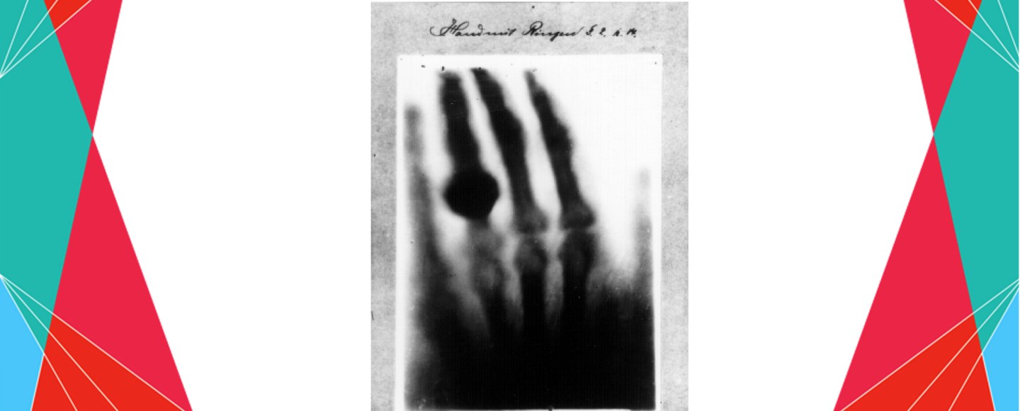


The invention of the x-ray machine marked a significant milestone in the medical world. On this day in 1896, scientist Henry Louis Smith formally exhibited the first x-ray machine. However, when he unveiled it, people were confused and unable to comprehend what he was trying to show them. It was only months later that German physics professor Wilhelm Röntgen discovered x-rays while conducting experiments with Lenard and Crookes tubes, leading to further research in the field.
The simplicity of an x-ray machine, such as those used in doctor’s or dentist’s offices, may come as a surprise. Essentially, it produces a controlled beam of radiation that is used to create an image of the inside of the body. Inside the machine is an x-ray tube, which contains an electron gun that shoots high-energy electrons at a target made of heavy atoms like tungsten.
In today’s world, getting an x-ray done is a relatively quick and easy process. However, the first x-ray machine required individuals to sit in front of it for over an hour to capture the necessary images. Additionally, the radiation dose applied to the subject was approximately 1,500 times higher than what is typically used in modern x-rays.
The introduction of the x-ray machine revolutionized medical diagnostics. Doctors and medical professionals could now easily visualize and identify even the smallest abnormalities within the body, aiding in accurate diagnoses and treatment plans. The ability to capture detailed images without invasive procedures allowed for a non-invasive approach, significantly reducing patient discomfort and improving overall healthcare outcomes.
Since the invention of the x-ray machine, it has become an indispensable tool in the medical field. It has evolved over the years, with advancements in technology leading to the development of more efficient and safer machines. Today, x-rays are used to diagnose various conditions, including bone fractures, lung infections, and even dental issues.
The impact of the x-ray machine on medical science cannot be overstated. Its ability to provide detailed images of the internal structures of the body has transformed healthcare practices and saved countless lives. Henry Louis Smith and Wilhelm Röntgen’s contributions to the field of radiology have paved the way for numerous advancements in medical imaging, benefiting patients worldwide.
#Hindi
एक्स-रे मशीन के आविष्कार ने चिकित्सा जगत में एक महत्वपूर्ण मील का पत्थर स्थापित किया। 1896 में, वैज्ञानिक हेनरी लुईस स्मिथ ने पहली बार एक्स-रे मशीन का प्रदर्शन किया। जब उन्होंने इसे दिखाया, तो लोग भ्रमित हो गए और समझ नहीं पाए कि वह क्या दिखाना चाह रहे थे। कुछ महीने बाद, जर्मनी के भौतिकी प्रोफेसर विल्हेम रॉन्टजन ने लीनार्ड और क्रुक्स ट्यूब का प्रयोग करते हुए एक्स-रे की खोज की, जिससे इस क्षेत्र में और अधिक शोध हुए।
एक्स-रे मशीन की सरलता देखकर हैरानी हो सकती है, जैसे डॉक्टर या दंत चिकित्सक के कार्यालय में उपयोग होने वाली मशीनें। मूल रूप से, यह नियंत्रित विकिरण की किरण बनाती है, जिसका उपयोग शरीर के अंदर की छवि बनाने के लिए किया जाता है। मशीन के अंदर एक एक्स-रे ट्यूब होती है, जिसमें एक इलेक्ट्रॉन गन होती है जो टंगस्टन जैसे भारी परमाणुओं के लक्ष्य पर उच्च ऊर्जा वाले इलेक्ट्रॉन फेंकती है।
आज के समय में, एक्स-रे कराना अपेक्षाकृत जल्दी और आसान प्रक्रिया है। लेकिन पहली एक्स-रे मशीन के लिए व्यक्ति को आवश्यक छवियाँ लेने के लिए एक घंटे से अधिक समय तक इसके सामने बैठना पड़ता था। इसके अलावा, उस समय प्रयुक्त विकिरण की मात्रा आधुनिक एक्स-रे की तुलना में लगभग 1,500 गुना अधिक थी।
एक्स-रे मशीन की शुरुआत ने चिकित्सा निदान में क्रांति ला दी। डॉक्टर और चिकित्सा पेशेवर अब शरीर के अंदर की छोटी से छोटी असमानताओं को आसानी से देख और पहचान सकते थे, जिससे सही निदान और उपचार योजनाओं में मदद मिली। बिना हस्तक्षेप किए विस्तृत छवियाँ लेने की क्षमता ने एक गैर-आक्रामक दृष्टिकोण की अनुमति दी, जिससे रोगी की असुविधा कम हुई और संपूर्ण स्वास्थ्य सेवा के परिणामों में सुधार हुआ।
एक्स-रे मशीन के आविष्कार के बाद से, यह चिकित्सा क्षेत्र में एक अनिवार्य उपकरण बन गया है। वर्षों में, प्रौद्योगिकी में प्रगति के साथ, अधिक कुशल और सुरक्षित मशीनें विकसित हुई हैं। आज, एक्स-रे का उपयोग हड्डी की टूटन, फेफड़ों के संक्रमण और यहां तक कि दंत समस्याओं जैसी विभिन्न स्थितियों का निदान करने के लिए किया जाता है।
चिकित्सा विज्ञान पर एक्स-रे मशीन का प्रभाव अत्यधिक महत्वपूर्ण है। इसके द्वारा शरीर की आंतरिक संरचनाओं की विस्तृत छवियाँ प्रदान करने की क्षमता ने स्वास्थ्य सेवा प्रथाओं को बदल दिया है और अनगिनत जिंदगियों को बचाया है। हेनरी लुईस स्मिथ और विल्हेम रॉन्टजन के रेडियोलॉजी के क्षेत्र में योगदान ने चिकित्सा छायाचित्रण में कई प्रगति के लिए मार्ग प्रशस्त किया है, जिससे विश्वभर के मरीजों को लाभ हो रहा है।
Share this: